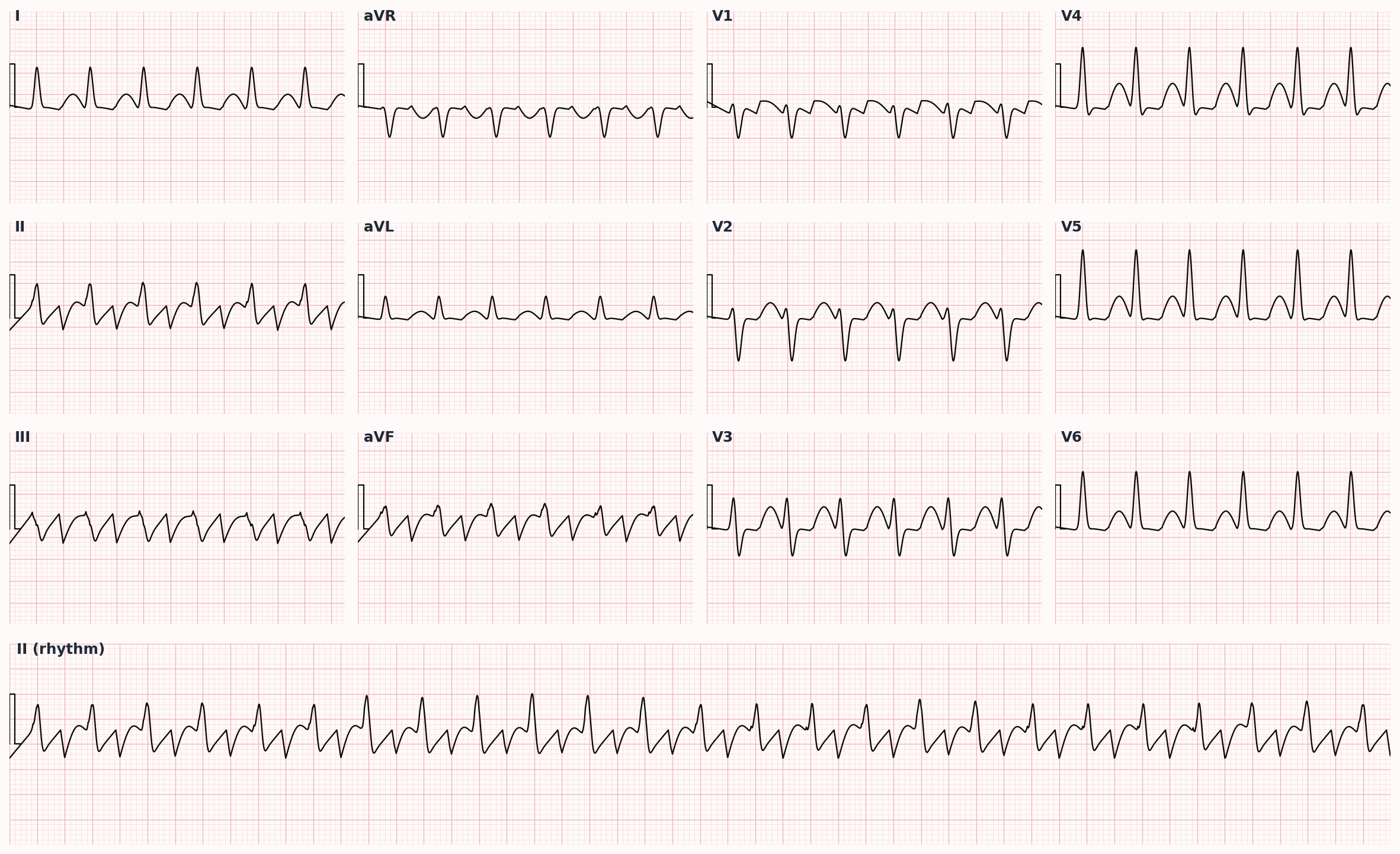
Macro-reentrant atrial tachycardia with sawtooth flutter waves; treated like AFib for stroke risk.
Also known as: AFlutter, flutter, atrial flutter, sawtooth, cavotricuspid isthmus
Overview
Organized macro-reentrant atrial tachycardia, typically with atrial rates 250-350 bpm (usually ~300), producing characteristic sawtooth flutter waves on ECG. Ventricular response depends on AV node conduction ratio (2:1, 3:1, 4:1).
Try two board-style Atrial Flutter questions
Real questions from the FirstPassPA bank, with the full explanation. Pick an answer — no signup, no email.
Question 1CardiovascularMedium
A 60-year-old man has newly recognized atrial flutter with an uncertain time of onset. His ventricular rate is adequately controlled and he is hemodynamically stable. His CHA₂DS₂-VASc score is 3, and he has not yet been anticoagulated. He wishes to undergo elective cardioversion to restore sinus rhythm. Which of the following is the most appropriate timing for elective cardioversion?
AImmediately, without any further evaluation
BAfter 1 week of therapeutic anticoagulation
CAfter bridging with low-molecular-weight heparin
DAfter ≥3 weeks of therapeutic anticoagulation
Reveal answer & full explanation
Correct answer: D — After ≥3 weeks of therapeutic anticoagulation
AImmediately, without any further evaluation
BAfter 1 week of therapeutic anticoagulation
CAfter bridging with low-molecular-weight heparin
DAfter ≥3 weeks of therapeutic anticoagulation✓
Why After ≥3 weeks of therapeutic anticoagulation is correct
For atrial fibrillation or flutter of ≥48 hours or unknown duration, elective cardioversion requires either ≥3 weeks of uninterrupted therapeutic anticoagulation beforehand or a transesophageal echocardiogram excluding left atrial appendage thrombus, per current AHA/ACC/HRS guidance
This patient is not yet anticoagulated and onset is uncertain, so the appendage cannot be assumed clot-free
Anticoagulation must also continue ≥4 weeks after cardioversion regardless of CHA₂DS₂-VASc score because of atrial stunning
Why the others are wrong
Immediately, without any further evaluation — cardioverting an un-anticoagulated patient with unknown-duration arrhythmia risks dislodging a pre-existing appendage thrombus (anchoring on hemodynamic stability while ignoring stroke risk)
After 1 week of therapeutic anticoagulation — one week is too short to organize or resolve an existing appendage thrombus; the guideline threshold is 3 uninterrupted weeks, or a negative TEE if earlier cardioversion is desired (right-idea-wrong-duration)
After bridging with low-molecular-weight heparin — heparin bridging is not the guideline pathway for elective cardioversion; the requirement is 3 weeks of therapeutic anticoagulation or a negative TEE (right-idea-wrong-step)
Question 2CardiovascularEasy
A 48-year-old woman presents with palpitations and is hemodynamically stable. The ECG shows a narrow-complex rhythm with an irregular ventricular response at approximately 110/min, no discernible P waves, and a regular sawtooth baseline undulating at an atrial rate of about 300/min that is most prominent in the inferior leads. Which of the following is the most likely diagnosis?
AAtrial flutter
BMultifocal atrial tachycardia
CAtrial fibrillation
DAtrioventricular nodal reentrant tachycardia
Reveal answer & full explanation
Correct answer: A — Atrial flutter
AAtrial flutter✓
BMultifocal atrial tachycardia
CAtrial fibrillation
DAtrioventricular nodal reentrant tachycardia
Why atrial flutter is correct
A regular sawtooth baseline at an atrial rate near 300/min, best seen in the inferior leads, is the signature of typical atrial flutter generated by a macro-reentrant circuit around the cavotricuspid isthmus.
Variable AV conduction (here roughly 3:1) produces an irregular ventricular response near 110/min, while the organized flutter waves remain visible between QRS complexes.
Recognition rests on these organized atrial waves rather than chaotic or absent atrial activity.
Why the others are wrong
Multifocal atrial tachycardia — requires at least three distinct P-wave morphologies with varying PR intervals; the absence of discrete P waves and the 300/min sawtooth exclude it (buzzword-match to 'irregular narrow-complex').
Atrial fibrillation — also irregular with no P waves, but shows chaotic fibrillatory undulations, not an organized sawtooth at a fixed 300/min atrial rate (the main premature-closure trap for any irregular SVT).
Atrioventricular nodal reentrant tachycardia — a regular narrow-complex tachycardia, usually 150-250/min, without flutter waves; the irregularity and sawtooth pattern here exclude it (anchoring on 'palpitations plus narrow complex').
🔒 Free preview limit reached
Keep reading — start your free trial
You've read your 2 free diagnosis previews. Create your free account to unlock the full Atrial Flutter outline — plus all 514 diagnoses, 6,500+ board-style questions, flashcards, and an AI tutor. Your 7-day free trial includes everything, and there's no credit card required.
Typical (counterclockwise) flutter: re-entrant circuit travels counterclockwise around the tricuspid annulus, with critical conduction through the cavotricuspid isthmus (CTI) at the floor of the right atrium. This anatomic substrate makes CTI ablation curative. Atypical flutter has variable circuits, often in the left atrium post-ablation or post-cardiac surgery.
Clinical presentation
Symptoms
Often more symptomatic than AFib due to faster, regular ventricular rates
Palpitations, dyspnea, exercise intolerance
Lightheadedness, chest discomfort
May present as new HF or with embolic stroke
Signs / physical exam
Regular tachycardia (in contrast to AFib's irregularly irregular)
Ventricular rate often a fixed multiple — 150 bpm (2:1), 100 (3:1), 75 (4:1)
Cannon a waves may be seen in JVP
Carotid massage or adenosine transiently blocks AV node — unmasks flutter waves diagnostically
Classic findings
ECG: 'sawtooth' flutter waves (negative deflections in II, III, aVF and positive in V1) in typical counterclockwise flutter.
Differential diagnosis
Atrial fibrillation (rate-controlled) — AFib at controlled rates can appear regular; absent P waves and fibrillatory baseline distinguish from flutter waves
AV nodal reentrant tachycardia (AVNRT) — Narrow-complex regular tachycardia, abrupt onset/offset, often 150-220 bpm; vagal maneuvers or adenosine terminate; pseudo-R' in V1 from retrograde P
AV reentrant tachycardia (AVRT, e.g., WPW) — Narrow-complex (orthodromic) or wide-complex (antidromic); delta wave visible in baseline sinus rhythm; AVOID AV-nodal blockers if pre-excited AFib
Sinus tachycardia at high rate — Gradual onset, identifiable trigger (fever, hypovolemia, anxiety, anemia), visible P waves with normal morphology
Focal atrial tachycardia — Single ectopic P-wave morphology different from sinus; rate 130-180 typically; less rapid than flutter
Ventricular tachycardia — Wide-complex; AV dissociation, capture/fusion beats, RBBB-like morphology >140 ms; assume VT until proven otherwise in patient with structural heart disease
Diagnostic workup
Labs
TSH, CBC, BMP, magnesium
Coagulation studies
Troponin if ischemia in differential
Imaging
12-lead ECG — diagnostic when flutter waves visible; 2:1 conduction can hide them (rate 150 should prompt vagal maneuvers or adenosine to unmask)
TTE for LA size, LV function, valve disease
TEE prior to cardioversion if duration unknown/>48 h and not adequately anticoagulated
Diagnostic algorithm
Feature
Atrial Fibrillation
Atrial Flutter
Atrial rate
350-600 bpm (chaotic)
250-350 bpm (~300 typical)
Ventricular rhythm
Irregularly irregular
Regular (or regularly irregular with variable block)
P waves
Absent — fibrillatory baseline
Sawtooth flutter waves (II, III, aVF negative in typical)
Typical ventricular rate
100-160 (variable)
150 (2:1), 100 (3:1), 75 (4:1)
Anticoagulation
CHA2DS2-VASc-driven
Same — equivalent stroke risk
Cardioversion energy
120-200 J biphasic
50-100 J biphasic
Curative ablation
PVI — ~70-80% success
CTI ablation — >90% success
AFib often follows ablation?
—
Yes (~30% over years)
Atrial flutter vs atrial fibrillation — board-relevant comparison. Stroke risk is identical; ablation success differs markedly.
Treatment
First-line
Acute rate control: same as AFib — IV beta-blocker or non-DHP CCB
Anticoagulation: same CHA2DS2-VASc-based decision as AFib (flutter carries equivalent stroke risk)
Cardioversion (synchronized DCCV) — flutter typically cardioverts at lower energy (50-100 J biphasic) than AFib (120-200 J)
Definitive: catheter ablation of cavotricuspid isthmus is curative for typical flutter with >90% success and low recurrence — preferred first-line in many patients
Second-line / adjunct
Antiarrhythmics (less effective than for AFib): ibutilide acutely; dofetilide, sotalol, amiodarone for maintenance
If cardioversion repeatedly fails or recurs despite AAD, refer for ablation
AFib commonly emerges after flutter ablation (~30% over years) — counsel patients and continue anticoagulation per CHA2DS2-VASc regardless of rhythm status
Complications
Embolic stroke (equivalent risk to AFib)
Tachycardia-induced cardiomyopathy (rapid 1:1 or 2:1 conduction sustained)
Heart failure
1:1 conduction with class IC antiarrhythmic (flecainide) — paradoxical rate acceleration; always pair flecainide with an AV nodal blocker
PANCE pearls
Regular narrow-complex tachycardia at exactly 150 bpm is atrial flutter with 2:1 conduction until proven otherwise. Use vagal maneuvers or adenosine to unmask the flutter waves diagnostically (not therapeutically).
CTI ablation has the highest success rate of any cardiac ablation procedure — counsel as first-line for most patients with typical flutter.
Apply the same stroke-prevention rules as AFib — flutter is NOT lower-risk for embolism despite being a more organized rhythm.
Atypical flutter often arises from prior left atrial ablation scars (post-PVI for AFib) — these require complex 3D mapping for ablation.
Images
Atrial flutter — sawtooth flutter waves (negative in II, III, aVF) with regular ventricular response
References
ACC/AHA/HRS 2015 SVT — 2015 ACC/AHA/HRS Guideline for the Management of Adult Patients with Supraventricular Tachycardia (Page et al., Circulation 2016)
ACC/AHA/ACCP/HRS 2023 — 2023 AFib Guideline applies same anticoagulation principles to flutter (Joglar et al., Circulation 2024)
Cosío 2017 — Atrial Flutter, Typical and Atypical: A Review (Cosío, Arrhythm Electrophysiol Rev 2017)
Practice Cardiovascular questions on FirstPassPA
Turn this outline into retention. 6,500+ board-style questions with an AI tutor that explains every answer — free to start, no card required.
Educational use only. This outline is a study aid for PA students and is not medical advice or a substitute for clinical judgment. FirstPassPA is an independent study tool and is not affiliated with, endorsed by, or sponsored by NCCPA or PAEA. PANCE® and PANRE® are registered trademarks of the National Commission on Certification of Physician Assistants; End of Rotation™ is a program of the Physician Assistant Education Association.